Epidemiology of Thyroglossal Duct Cysts in an Eastern Caribbean Nation
Gailann Jugmohansingh, Steve Medford, Shariful Islam
DOI10.21767/2572-2107.100002
Jugmohansingh G1, Medford S1 and Islam S2*
Department of Otorhinolaryngology, Head and Neck Surgery, San Fernando General Hospital, Trinidad and Tobago
San Fernando General Hospital, Trinidad and Tobago
- *Corresponding Author:
- Shariful Islam
DM (General Surgery)
San Fernando General Hospital
Trinidad and Tobago
Tel: 868-797-4951
E-mail: sssl201198@yahoo.com
Received date: Oct 01, 2015; Accepted date: Dec 02, 2015; Published date: Dec 10, 2015
Citation: Jugmohansingh G1, Medford S, Islam S. Epidemiology of Thyroglossal Duct Cysts in an Eastern Caribbean Nation. Head Neck Cancer Res. 2015, 1:1.
Abstract
Aim: The thyroglossal duct cyst is the most common congenital cystic lesion in the neck. Seven percent of the population has persistence of this duct. It usually occurs in midline and is asymptomatic. The aim of this study is to see the epidemiology of this disease in an eastern Caribbean nation.
Design and methods: An 11 year’s retrospective study was performed at the San Fernando General Hospital from 1st January 2004 to 31st December 2014. All of patients who had a thyroglossal duct cyst excision from the surgical departments were reviewed. 17 patients were identified. Data on gender, ethnicity, age, reason for seeking medical attention and location of the cyst were extracted.
Results: M:F was 7:10, Ethnic distribution: East Indians 65% and Africans 35%, age range birth 51 years, symptomatic patients 65% (unique description of an infected right lateral thyroglossal duct cyst with suprahyoid, anterior hyoid and infrahyoid components), location: midline 71% and right lateral 29% with no left lateral cases.
Conclusions: Thyroglossal duct cysts at the San Fernando General Hospital have a mild female preponderance, range from birth to 51 years, is clinically evident soon after head and neck infection and have a higher occurrence of right lateral cysts at the suprahyoid level. An increased number of symptomatic and right lateral thyroglossal cysts were also noted in our population.
Keywords
Thyroglossal duct cyst; Neck mass; Sistrunk procedure; Sclerotherapy
Introduction
Cystic neck abnormalities can be broadly classified into congenital and acquired lesions. There are multiple diagnoses for these lesions but the thyroglossal duct cyst accounts for the majority of cases [1-3].
The diagnosis of a thyroglossal duct cyst is made after an appropriate history, physical examination and relevant radiological investigations. The standard treatment of choice is surgical removal following the Sistrunk procedure [4,5]. Only recently, sclerotherapy with OK 432 has become an alternative in selected cases [6,7].
In the past few years at the San Fernando General hospital, we have seen thyroglossal duct cysts with unusual presentations and in unusual locations. This has caused misdiagnosis and led to poor management in some cases. Due to these concerns, a retrospective study on thyroglossal duct cysts was undertaken to document how affected patients presented and also to document the location of the cysts.
Methods
An 11 year retrospective case series was conducted at the tertiary institution of the San Fernando General Hospital from January 1st 2004 – December 31st 2014. The operative logbooks of Otorhinolaryngology, Pediatric surgery and General surgery were examined and all patients who underwent excision of a thyroglossal duct cyst (initial or subsequent procedure) were documented.
The notes of these patients were then obtained through the Medical records department. The sole inclusion criteria for the study were that a thyroglossal duct excision/ removal or Sistrunk procedure had to be documented in the operative logbooks. The exclusion criteria were: no notes being recovered, no operative notes in recovered file and/or the final histology not confirming a thyroglossal duct cyst.
Information about gender, ethnicity, age, reason for seeking medical attention and location of the cyst (based on clinical, ultrasound and operative findings) were extracted from each file. The results were then analysed and compared to the international literature.
Results
Twenty five cases of thyroglossal duct cyst excisions were documented in the operative logbooks. However, eight cases had to be excluded. The notes for three cases were unable to be recovered, no operative notes were found in two files and three histologies documented histologies other than a thyroglossal duct cyst.
Of the seventeen confirmed cases, the departments of Otorhinolaryngology, Pediatric surgery and General Surgery managed 76.47%, 11.76% and 11.76% of patients respectively.
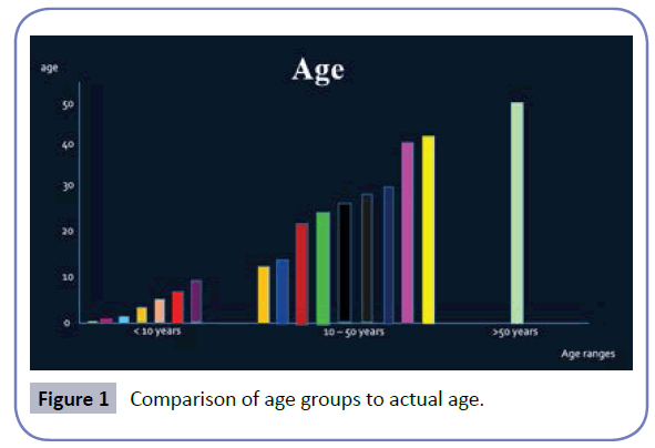
The male to female ratio was 7:10. East Indians accounted for 65% of cases while Africans made up the remaining 35% (Figure 1).
The ages ranged from birth to 51 years old. With respect to age, 2 peaks were noted at 5 – 10 years and 20 – 30 years. Below the age of ten, seven people had thyroglossal duct cysts. Within the age group 10 – 50, 9 patients had cysts. In this series, only one patient developed a cyst over the age of fifty.
The clinical presentation of the patients fell into one of five categories: infected cyst, asymptomatic, dysphagia/ shortness of breath, discharging sinus and a painful mass with rapid increase in size and compressive features. An infected cyst was the most common presentation (Table 1).
| Clinical presentation | Number of patients |
|---|---|
| Asymptomatic Cases | 6 |
| Infected Cysts | 7 |
| Discharging sinus | 1 |
| Dysphagea/intermittent dyspnea | 2 |
| Painful, rapid increase in size + compression | 1 |
Table 1: Different clinical presentations of a thyroglossal duct cyst.
Midline thyroglossal duct cysts represented 71% of all cases. Right sided cysts accounted for 29% of all cases. There were no documented cases of left sided cysts. Within the midline thyroglossal duct cyst group, suprahyoid and thyrohyoid cysts occurred with frequencies of 7% and 60% respectively. There were no cases of intralingual or suprasternal cysts (Table 2). In the right sided cystic group, the suprahyoid level was the most common site (Table 3).
| Location | Percentages |
|---|---|
| Intra-lingual | 0 |
| Suprahyoid | 8 |
| Thyrohyoid | 92 |
| Suprasternal | 0 |
Table 2: The distribution of midline cysts.
| Location | Percentages |
|---|---|
| Infrahyoid | 20 |
| Hyoid level | 0 |
| Suprahyoid | 60 |
| *All levels | 20 |
Table 3: The distribution of lateral cysts.
Discussion
Cystic lesions of the neck include branchial cleft cysts, thyroglossal duct cysts, lymphangioma, dermoid cysts, epidermoid cysts, infections/inflammatory masses, cystic lesions of the thyroid, thymic cysts, laryngoceles, ranulas, cystic lesions of salivary glands, cystic metastatic lymph nodes, neurogenic tumours, rare vascular lesions and cervical bronchogenic cysts [8-10]. The thyroglossal duct cyst accounts for 70% of all congenital neck masses [1-3].
The thyroid gland descends from the base of the tongue at the level of the foramen caecum into the anterior neck through the thyroglossal duct [11,12]. This duct usually undergoes involution but in seven percent of patients it remains patent [1,13]. The ducts have an epithelial lining which - in the presence of infection or inflammation produces excess secretions that cause ductal dilatation and cyst formation [14,15]. The patient may seek medical attention for cosmetic concerns regarding an asymptomatic cyst or when the cyst becomes symptomatic.
In this small case series, the statistics reported in the age groups less than fifty years compared favorably to international statistics [2,16]. Ahura et al. and Fischer et al. also reported a greater percentage of patients over fifty years that developed cysts. No particular reason was found to account for this.
The M:F ratio was 7:10. Internationally no gender preference has been documented [17,18]. However in a recent review of 159 cases by Taimisto et al. a mild female predominance was noted [19]. Our case series also reports a mild female predominance.
The population of Trinidad and Tobago is divided as follows: East Indians: 40%, Africans: 37.5%, Mixed ethnicity: 20.5%, Others: 1.2% and Unspecified: 0.8% [20]. There appears to be a higher number of East Indians with thyroglossal duct cysts. However due to the small number of patients, it cannot be said with any certainty that this ethnic group is predisposed to cyst formation (Table 4).
| Age Range | International % (Ahura et al 2005, Fischer et al 2007) | SFGH % |
|---|---|---|
| <10 years | 40 | 41 |
| 10 - 15 years | 45 | 53 |
| >50 years | 15 | 6 |
Table 4: Age of patients at SFGH compared to international statistics.
The population of Trinidad and Tobago is divided as follows: East Indians: 40%, Africans: 37.5%, Mixed ethnicity: 20.5%, Others: 1.2% and Unspecified: 0.8% [20]. There appears to be a higher number of East Indians with thyroglossal duct cysts. However due to the small number of patients, it cannot be said with any certainty that this ethnic group is predisposed to cyst formation.
Patients with clinically evident thyroglossal duct cysts can be either asymptomatic or symptomatic. The literature has described cysts as being largely asymptomatic [14,21-23]. However, in this study, only 35% of patients were asymptomatic. The majority of patients were actually symptomatic and accounted for 65% of all cases. Notably, these patients all had head or neck infections before presentation. Six patients had a recent upper respiratory tract infection, one patient had cellulitis of the anterior neck and one patient had an infected cyst that developed a discharging fistula. The inflammation associated with infection could have increased secretions of the epithelium within the thyroglossal duct producing an obvious cystic swelling in the neck (Table 5) [14,15].
| Location | International (Shah et al 2005, Ghaneim et al 2007, Allard 1982) | SFGH | |
|---|---|---|---|
| Intra-lingual | 2% | 0% | |
| Suprahyoid | 24% | 6% | |
| Thyrohyoid | 60% | 65% | |
| Suparsternal | 13% | 0% | |
Table 5: Midline cysts – International vs SFGH statistics.
Within the subgroup of midline cysts, the thyrohyoid level was the most common location internationally and locally [3,14,24]. However, suprahyoid cysts only occurred in 6% of patients at the SFGH. There were no cases of intralingual or suprasternal cysts. A larger sample size may have included the latter two types.
Internationally, ten percent of patients have been reported to have lateral thyroglossal duct cysts [25-27]. The findings of this study therefore represents almost three times the normal occurrence. The suprahyoid location of the lateral cyst was the lowest international percentage published [27]. However, it was the most common level for occurrence in this series. There was also the description of a unique entity, a thyroglossal duct cyst that had suprahyoid, hyoid and infrahyoid components. Therefore, the low degree of suspicion for thyroglossal duct cysts in these lateral masses may be the reason why many of the cysts were being misdiagnosed as other entities at the San Fernando General Hospital (Table 6).
| Location | Internationally (Ahuja et al 1999) | SFGH |
|---|---|---|
| Infrahyoid | 26-65% | 20% |
| Hyoid level | 15-50% | 0% |
| Suprahyoid | 20-25% | 60% |
| *All levels | Not described | 20% |
Table 6: Lateral cysts: International vs SFGH statistics.
Conclusion
There appears to be a mild female preponderance in patients with thyroglossal duct cysts at the San Fernando General Hospital. The ages of patients presenting to the hospital range from birth to 51 years. The majority of patients were symptomatic and cysts seemed to develop soon after head and neck infections. There is an increased incidence of right lateral thyroglossal duct cysts found at the suprahyoid level and a high degree of suspicion is necessary for accurate diagnosis of this entity.
Disclosure
The authors have nothing to disclose.
Acknowledgements
1. Dr M. Ashraph, Dr. R. Maharaj, Dr N. Armoogum- From the Department of Otorhinolaryngology - San Fernando General Hospital, Trinidad and Tobago
2. Dr. L. Roop, Dr. R Persad- From the Department of Pediatric Surgery - San Fernando General Hospital, Trinidad and Tobago
3. Dr V. Bheem, Dr S. Budhooram, Dr J Shah, Dr D. Dan, Dr. K. Sookhoo, Dr. T. Kuruvilla and Dr. Y. Maharaj- From the Department of General Surgery - San Fernando General Hospital, Trinidad and Tobago
References
- Ellis PD, Van Nostrand AW (1977) The applied anatomy of thyroglossal tract remnants. The Laryngoscope 87: 765-770.
- Ahuja AT, Wong KT, King AD, Yuen EHY (2005) Imaging for thyroglossal duct cyst: the bare essentials. Clinical radiology 60: 141-148.
- Allard RH (1982) The thyroglossal cyst. Head & neck surgery 5: 134-146.
- Sistrunk WE (1920) The surgical treatment of cysts of the thyroglossal tract. Annals of surgery 71: 121.
- Brunicardi FC, Anderson DK, Billiar TR, Dunn DL, Hunter JG, Pollock RE (2005) Pediatric Surgery. In : Shwartz’s principles of surgery. McGraw-Hill, New York.
- Kim MG, Kim SG, Lee JH, Eun YG, Yeo SG (2008) The Therapeutic Effect of OK-432 (Picibanil) Sclerotherapy for Benign Neck Cysts. The Laryngoscope 118: 2177-2181.
- Ohta N, Fukase S, Suzuki Y, Ishida A, Aoyagi M (2010) Treatments of various otolaryngological cystic diseases by OK-4321. The Laryngoscope 120: 2193-2196.
- Mittal MK, Malik A, Sureka B, Thukral BB (2012) Cystic masses of neck: A pictorial review. The Indian journal of radiology & imaging 22: 334.
- Saylam G, Tanrikulu S, Dursun E, Iriz A, Eryilmaz A (2008) A mass at fat density in the parotid gland: dermoid cyst or lipoma. B-ENT 5: 43-45.
- Al-Khateeb TH, Al Zoubi F (2007) Congenital neck masses: a descriptive retrospective study of 252 cases. Journal of Oral and Maxillofacial Surgery 65: 2242-2247.
- Moore KL, Persaud TVN (2003) The pharyngeal apparatus. In: The developing human: Clinically oriented embryology 7th edn Saunders, Philadelphia.
- Schoenwolf GC, Bleyl SB, Brauer PR, Francis-West PH (2009) Development of the pharyngeal apparatus and face. In: Larsen’s human embryology, 4th edn Churchill Livingston Elsevier, Philadelphia.
- Simon LM, Magit AE (2012) Impact of incision and drainage of infected thyroglossal duct cyst on recurrence after Sistrunk procedure. Archives of Otolaryngology–Head & Neck Surgery 138: 20-24.
- Shah R, Gow K, Sobol SE (2007) Outcome of thyroglossal duct cyst excision is independent of presenting age or symptomatology. International journal of pediatric otorhinolaryngology 71: 1731-1735.
- Shvili I, Hadar T, Sadov R, Koren R, Shvero J (2009) Cholesterol granuloma in thyroglossal cysts: a clinicopathological study. European Archives of Oto-Rhino-Laryngology 266: 1775-1779.
- Fischer JE, Bland KI (2007) Congenital lesions: thyroglossal duct cyst, branchial cleft anomalies and cystic hygromas. In: Mastery of surgery 5th edn. Lippincott Williams & Wilkins, Philadelphia.
- Brousseau VJ, Solares CA, Xu M, Krakovitz P, Koltai PJ (2003) Thyroglossal duct cysts: presentation and management in children versus adults. International journal of pediatric otorhinolaryngology 67: 1285-1290.
- Galluzzi F, Pignataro L, Gaini RM, Hartley B, Garavello W (2013) Risk of recurrence in children operated for thyroglossal duct cysts: a systematic review. Journal of pediatric surgery 48: 222-227.
- Taimisto I, Mäkitie A, Arola J, Klockars T (2015) Thyroglossal duct cyst: patient demographics and surgical outcome of 159 primary operations. Clinical Otolaryngology 40: 496-499.
- https://www.indexmundi.com/trinidad_and_tobago/demographics_profile.html
- Brewis C, Mahadevan M, Bailey CM, Drake DP (2000) Investigation and treatment of thyroglossal cysts in children. Journal of the Royal Society of Medicine 93: 18-21.
- Lin ST, Tseng FY, Hsu CJ, Yeh TH, Chen YS (2008) Thyroglossal duct cyst: a comparison between children and adults. American journal of otolaryngology 29: 83-87.
- Ostlie DJ, Burjonrappa SC, Snyder CL, Watts J, Murphy JP, et al. (2004) Thyroglossal duct infections and surgical outcomes. Journal of pediatric surgery 39: 396-399.
- Ghaneim A, Atkins P (1996) The management of thyroglossal duct cysts. International journal of clinical practice 51: 512-513.
- Latham K, Perry WB, Richards ML (2006) Lateral Neck Mass in a Young Woman. Current surgery 63: 24-26.
- Acierno SP, Waldhausen JH (2007) Congenital cervical cysts, sinuses and fistulae. Otolaryngologic Clinics of North America 40: 161-176.
- Ahuja AT, King AD, King W, Metreweli C (1999) Thyroglossal duct cysts: sonographic appearances in adults. American journal of neuroradiology 20: 579-582.
Open Access Journals
- Aquaculture & Veterinary Science
- Chemistry & Chemical Sciences
- Clinical Sciences
- Engineering
- General Science
- Genetics & Molecular Biology
- Health Care & Nursing
- Immunology & Microbiology
- Materials Science
- Mathematics & Physics
- Medical Sciences
- Neurology & Psychiatry
- Oncology & Cancer Science
- Pharmaceutical Sciences

