Pleomorphic Adenoma of Ectopic Salivary Gland Tissue in the Upper Neck
Chang CF and Wang CW
DOI10.21767/2572-2107.100009
1Department of Otorhinolaryngology, Head and Neck Surgery, Jen-Ai Hospital, Taichung, Taiwan
2Central Taiwan University of Science and Technology, Taichung, Taiwan
3School of Medicine, Chung Shan Medical University, Taichung, Taiwan
4Department of Otorhinolaryngology, Head and Neck Surgery, Changhua Christian Hospital, Changhua, Taiwan
- *Corresponding Author:
- Chang CF
Department of Otorhinolaryngology
Head and Neck Surgery
Jen-Ai Hospital
Taichung, Taiwan
Tel: 886424819900
E-mail: benglung@hotmail.com
Received date: June 20, 2016; Accepted date: July 15, 2016; Published date: July 22, 2016
Citation: Chang CF, Wang CW. Pleomorphic Adenoma of Ectopic Salivary Gland Tissue in the Upper Neck. Head Neck Cancer Res. 2016, 1:2.
Keywords
Barriers to cessation; Reasons for quitting; Cigarette; Drinking; Concurrent
Introduction
Pleomorphic adenoma also known as “mixed tumor” is the most common neoplasm in major salivary glands. It is a benign tumor arises from the parotid gland in 84% of cases and represents 45% of parotid gland tumors [1]. Only 6.5% are found in the minor salivary glands [2]. Salivary gland tumors occurring in ectopic sites are rare and poorly understood; sometime we can find heterotopic tissue arising from an aberrant salivary gland in external auditory canal, nasal cavity, tongue, tonsils and neck [3]. The presence of pleomorphic adenoma in the upper neck, along the anterior border of the sternocleidomastoid muscle is extremely rare. Only five cases of ectopic mixed salivary gland tumors in the upper neck in adult patients have been reported [4-7]. This article describes another case of pleomorphic adenoma occurring in the upper neck followed by a literature review in order to identify the major characteristics and differential diagnosis of such a rare disease.
Case Report
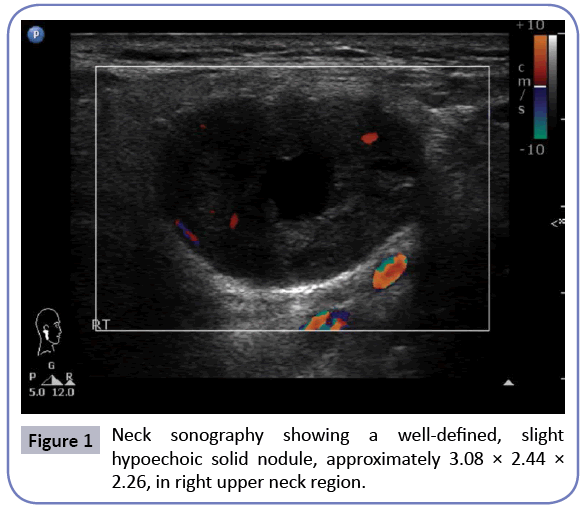
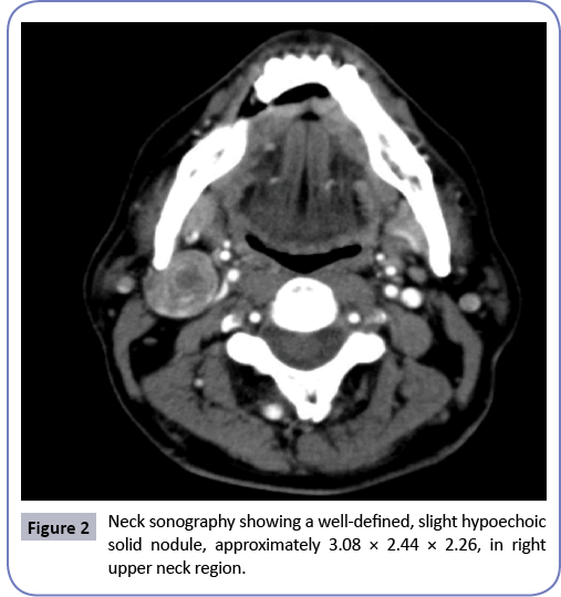
A 59 year old man presented as an outdoor patient complained of a painless mass in the right neck for approximately 3 months. There were no other symptoms, no clinical evidence or family history. In a physical examination the mass was found to be located in the right level II, below the right mandible angle, lying on the anterior border of the right sternocleidomastoid muscle. An ultrasound scan of the neck showed a hypoechoic, welldefined nodule measuring about 3.08 × 2.44 × 2.26 cm in size in the right submandibular area (Figure 1). Fine-needle aspiration cytology (FNAC) of the lesion reported no presence of malignant cells. In a CT scan of the neck we found a 2.43 cm heterogeneously enhancing nodular lesion with well-defined margins in right level II (Figure 2). No invasion into the surrounding tissues was observed. The lesion was completely excised under general anesthesia.
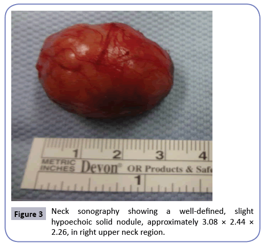
The surgical specimen consists roughly of one brownish elastic tissue fragment measuring 3.2 × 2.4 × 1.8 cm in size and in a fresh state (Figure 3). Histologic examination revealed a well demarcated nodule consisting of proliferative epithelial cells and myoepithelial cells with focal ductal differentiation and myxoid to chondroid matrix in focal areas (Figure 4). The immunohistochemical study demonstrates CK7 (+) in the epithelial cells and P63 (+) in the myoepithelial cells. The CD117 stain shows focal weak expression. The Ki-67 labeling index is very low. Scant salivary gland tissues are seen in the peripheral area. In conclusion, pleomorphic adenoma (mixed tumor) is considered.
The post-surgical course was normal and the patient was discharged two days later. There is no sign of local or distant recurrence in six months after first operation.
Discussion
Pleomorphic adenoma originates from the epithelial and myoepithelial cells of the intercalar ducts and is characterized histologically by different types of tissues (glandular, epithelial, myoepithelial, myxoid, fibrous, chondral, and bony) [8]. It is the most common type of salivary gland tumor and the most common tumor in the parotid gland. The tumor is usually solitary and presents as a slow growing, painless, firm single nodular mass. The high cellularity and solid character of pleomorphic adenoma usually leads to misdiagnosis as more aggressive neoplasms. For this reason, histologic examination of the entire specimen is suggested. The potential risk of malignant transformation of the pleomorphic adenoma is about 6%, predominantly seen in female patients [9], and is increased by delay in diagnosis.
Typical locations of pleomorphic adenoma have been described to be in small salivary glands, accessory glands, or ectopic sites. Salivary tissues found in unusual locations is termed ectopic or heterotopic salivary tissues, as well as salivary tissue choristoma [10]. Salivary glands have been described to be in a variety of aberrant locations, including the hypophysis, cerebellopontile angle, middle ear, mastoid, auditory canal, tongue, palatine tonsil, thyroglossal duct, mandible, thyroid and parathyroid capsules, and sternoclavicular joints [3] In our literature review, the first description of ectopic salivary glands was recorded in 1789 [5]. Many theories have been put forward to explain the occurrence of this condition: abnormal persistence and development of vestigial structures, dislocation of a portion of definitive organ rudiment mass, and further development along with abnormal differentiation of local tissues. Now days, the best accepted theory is that the presence of heterotopic salivary tissues in the neck arises from epithelial remnants in the branchial apparatus [7,11-15].
Many papers have reported the presence of salivary heterotopic tissues in the neck, but the presence of salivary ectopic tissues in the upper neck area is extremely rare. Following a literature review, we found only five previous adult cases of pleomorphic adenoma in the upper neck. The first case of the tumor occurring in the neck was described by Pesavento and Ferlito [4] two years later Hulbert published the second case [5] followed by the third one reported by Ordonez et al. [6]. Domenico et al. reported the fourth case in 2008 [7] and in 2012 LuksiÃÆââ¬Å¾Ãâââ¬Â¡ et al. presented the fifth one [14].
Differential diagnosis of masses in the upper neck includes developmental anomalies (brachial cleft cyst, epidermoid cyst, thyroglossal duct cyst), infections, benign tumors (Warthin’s tumor, pleomorphic adenoma, neurogenic neoplasm), and malignant neoplasms (mucoepidermoid carcinoma, acinic cell carcinoma, anaplastic carcinoma, metastatic malignancy) [10,12,13]. In general, computed tomography (CT) of the neck is the most helpful test. It may differentiate solid masses from cystic masses, locate a mass within a glandular structure or identify it as a free nodal lesion, and differentiate congenital vascular lesions from lymph nodal chains. Fine-needle aspiration biopsy is useful in confirming clinical diagnosis of a cystic lesion and is appropriate before excision biopsy [12].
Mixed salivary tumors are the most frequent occurrences among ectopic neoplasm; they have the same percentage of recurrence but a higher percentage of cancerization than those occurring in major salivary glands [5,9,11]. Other tumors arising within ectopic salivary glands, within and beyond the confines of the neck, include adenolymphoma, adenoid cystic carcinoma, mucoepidermoid carcinoma, and anaplastic carcinoma [10,13]. Clinically, pleomorphic adenoma in the upper neck manifests as a lowly enlarging, painless and movable mass, while the lower cervical ectopic salivary tissue usually presents as a draining sinus [14]. The treatment for ectopic mixed salivary tumor endorsed by all authors is the complete surgical excision.
In the presence of a slow-growing unilateral mass in the upper neck, we should be careful with its diagnosis and treatment, which often present incidence of unexpected pathology to head and neck surgeons. Early diagnosis offers the possibility of a more complete excision with adequate care being taken not to disrupt the tumor in order to prevent local and distant spread of neoplastic cells. Long-term follow-up to exclude malignancy is mandatory, even if the tumor appears to be clinically benign and removed completely.
References
- Smolka W, Eggensperger N, Stauffer-Brauch EJ, von Bredow F,Lizuka T (2007) Pleomorphic adenoma in an atypical location near the temporomandibular joint: a case report. Quintessence Int. 38:417-421.
- Drinkard DW, Schow CE Jr (1986) Benign mixed tumor of the mandible 17 years after the occurrence of a similar lesion in the parotid gland. Oral Surg Oral Med Oral Pathol62:381-384.
- Ross DE,Sukis AE (1971) Salivary gland tumors in ectopic sites. Laryngoscope 81: 558-564.
- Pesavento G, Ferlito A (1976) Benign mixed tumour of heterotopic salivary gland tissue in upper neck. Report of a case with a review of the literature on heterotopic salivary gland tissue. J Laryngol Otol90: 577-584.
- Hulbert JC (1978) Ectopic mixed salivary tumour in the neck. J Laryngol Otol 92: 533-536.
- Ordonez MM, Sancho Alvarez A, Morais Perez D, Alvarez Gago T (1994) Mixed tumour in cervical salivary heterotopy. Acta OtorrinolaringolEsp 45: 387-389.
- DomenicoT (2008) Pleomorphic Adenoma in Ectopic Salivary Tissue of the Neck. Open Otorhinolaryngology Journal 2: 13.
- Youngs LA, Schofield HH (1976) Arch Pathol 83: 550.
- Shaheen OH (1997) Benign salivary gland tumours: Scott Brown’s Otolaryngology, 6th edn, Oxford, Butterworth-Heinemann, p: 5.
- Solem BS, Schroder KE, Mair IW (1981)Differential diagnosis of a mass in the upper lateral neck. J Laryngol Otol 95:1041-1047.
- Cotelingam JD, Gerberi MP (1983) Parotid heterotopia with pleomorphic adenoma. Arch Otolaryngol. 109: 563-565.
- Su CC, Chou CW, Yiu CY (2008)Neck mass with marked squamous metaplasia: a diagnostic pitfall in aspiration cytology. J Oral Pathol Med 37:56-58.
- Batsakis JG (1974) Tumors of the head and neck. Williams and Wilkins: Baltimora.
- LuksiÃÆââ¬Å¾Ãâââ¬Â¡ I, Suton P, ManojloviÃÆââ¬Å¾Ãâââ¬Â¡ S, Macan D, Dediol E (2012) Pleomorphic adenoma in ectopic salivary gland tissue in the neck. Coll Antropol. Suppl 2:133-136.
- Arunkumar KV, Kumar S, Bansal V, Saxena S, Elhence P (2011) Pleomorphic adenoma--unusual presentation of a salivary gland tumor in the neck of a child. Quintessence Int42:879-882.
Open Access Journals
- Aquaculture & Veterinary Science
- Chemistry & Chemical Sciences
- Clinical Sciences
- Engineering
- General Science
- Genetics & Molecular Biology
- Health Care & Nursing
- Immunology & Microbiology
- Materials Science
- Mathematics & Physics
- Medical Sciences
- Neurology & Psychiatry
- Oncology & Cancer Science
- Pharmaceutical Sciences


