Septal Chondroma that Cause Deviation of the Nose: Case Report
Wael Y Elias and Nizar Fageeh
DOI10.21767/2572-2107.100018
Nizar Fageeh1 and Wael Y Elias2*
1Oral Diagnostic Science Department, Faculty of Dentistry, King Abdulaziz University, Jeddah, Saudi Arabia
2Department of Otorhinolaryngology, King Fahd Hospital, Jeddah, Saudi Arabia
- *Corresponding Author:
- Elias WY
Oral Diagnostic Science Department, Faculty of Dentistry
King Abdulaziz University, Jeddah, Saudi Arabia
Tel: +966 12 640 1000
E-mail: dreliasw@gmail.com
Received date: 25 September 2017; Accepted date: 04 October 2017; Published date: 11 October 2017
Citation: Elias WY. Septal Chondroma that Cause Deviation of the Nose: Case Report. Head Neck Cancer Res. 2017, Vol.2 No.1:3. doi:10.21767/2572-2107.100018
Copyright: © 2017 Elias WY, et al. This is an open-access article distributed under the terms of the Creative Commons Attribution License, which permits unrestricted use, distribution, and reproduction in any medium, provided the original author and source are credited.
Abstract
Cartilaginous tumors of the nasal septum are extremely rare. We describe a 17-year-old male patient who presented with nasal deformity and complete obstruction of one of the nasal passages since childhood. The tumor was located anteroinferiorly on the nasal septum. A biopsy revealed findings consistent with chondroma of the nasal septum. The tumor was removed completely by a combined endoscopic and external subnasal approach. The patient was followed up and after two years, he did not show any signs of recurrence on re-evaluation.
Keywords
Cartilaginous tumors; Chondroma; Nasal septum
Introduction
Cartilage tumors of the head and neck are rare, especially those of the nasal septum. Nasal chondroma was first described [1]. Only about 131 cases were described [2]. It was found 50% of nasal chondromas originating from the ethmoid and only 17% from the nasal septum [3]. A review of the literature by Rivas et al. discovered only 19 chondromas arising from the nasal septum, almost always on the posterior part of the septum. We report a rare case of nasal septum chondroma in a 17-year-old teenager.
Case report
A 17 year old Saudi male patient was referred to our institution with a nasal deformity for rhinoplasty. The patient presented with nasal deformity and a history of complete obstruction of one of the nasal passages since childhood. There was no history of epistaxis, nasal allergy, postnasal discharge, sleep apnea, or nasal trauma. His history was also unremarkable for systemic disease, and he had no family history of diabetes or malignancy.
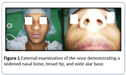
Physically, the patient appeared to be in good health, with a normal body weight. External examination of the nose showed a widened nasal bone, broad tip, nasolabial angle of 90°, and wide alar base (Figure 1).
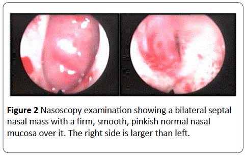
Anterior rhinoscopy revealed the nasal septum to be deviated to the right side (Figure 2).
A pinkish, bony, hard mass filled both sides of the nasal cavity and was covered with a normal appearing mucosa that could not be distinguished from the nasal septum. It was not possible to pass beyond the mass. The remainder of the head, neck, and physical examination was normal.
Contrast computed tomography scan of the paranasal sinuses revealed a mass arising from the inferior aspect of the nasal septum or left maxilla. The paranasal sinuses, orbital cavity, lamina papyracea, and cribriform plate were intact. The mass compressed the inferior and middle turbinate’s, particularly on the right side. Magnetic resonance imaging revealed higher signal intensity on T2W1 compared to a low to intermediate signal intensity on T1W1. A bone scan was normal.
Examination under general anesthesia confirmed the origin of the mass to be from the anteroinferior part of the septum. A biopsy of the mass showed benign tumor of cartilage consistent with septal chondroma.
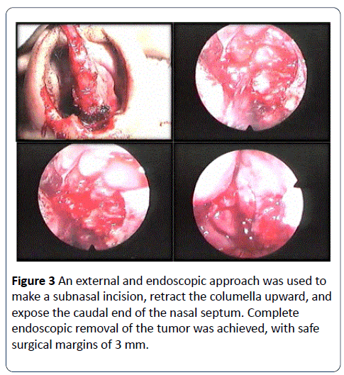
The tumor was removed under general anesthesia using a combined external and endoscopic technique (Figure 3).
The tumor was approached through a wide sub nasal incision, and the columella was retracted upward and left intact. The caudal septum was exposed, and a bilateral mucoperichondrial flap was raised, revealing 15 mm of the caudal end of the septum to be intact. Once the mass was exposed, fragmented chips were removed from the posterior lower quadrilateral cartilage of the septum. The anterior part of the ethmoid bone of the septum was removed. After removing a 3 mm margin of the normal septum, it was possible to leave 10 mm of the caudal and upper parts of the septum intact.
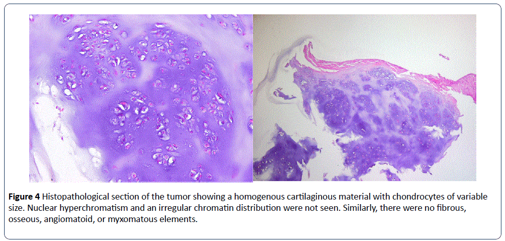
Histopathological examination of the excised tumor revealed a homogenous cartilaginous material with chondrocytes of variable size (Figure 4).
There was no evidence of anisochromia or hyperchromatic nuclear densities. Furthermore, fibrous, osseous, angiomatoid, or myxomatous elements were not present. The definitive diagnosis was benign chondroma of the nasal septum.
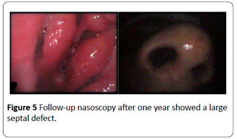
The patient was re-evaluated after two years, with endoscopic examination showing a large septal perforation (Figure 5).
There was no endoscopic evidence of any residual tumor, and the caudal and superior ends of the septum were still intact. The external appearance of the nose was unchanged without evidence of saddling, nasal tip projection, or change in the nasolabial angle. A biopsy from the edge of the septum was negative.
Discussion
Chondromas of the head and neck are rare, benign tumors that are most commonly found in the long bones, ribs, or pelvis [4,5]. When found in the head and neck region, they are commonly encountered in the maxillofacial area or larynx [6]. Septal chondroma is a very rare slow- growing tumor [2,3]. In this case, the patient presented with complete nasal obstruction and nasal deformity, which could have been mistaken for a severely deviated nasal septum because the mucosa remained intact.
The cell rest theory is the most accepted concept in the carcinogenesis of nasal chondroma, with chondrogenesis arising in the posterior area of the nasal septum, paranasal sinuses, turbinates, or the hard palate [7,8]. Trauma, metaplasia, and heredity may also play a role in the etiology of the tumor [8], but these were unlikely in the current case due to the absence of a history of trauma and the patient’s unremarkable family history.
Lateral rhinotomy has traditionally been the treatment of choice in patients with nasal chondroma. Various open rhinoplasty approaches have also been recommended [8,9]. Our decision was to use a combined subnasal and endoscopic approach, which permitted us to preserve the columella, completely expose the caudal end of the septum, and completely excise the tumor, leaving tumor-free margins. The upper and caudal septal cartilage was preserved as much as possible to maintain the shape of the nose.
Nasal septal chondroma should be considered in the differential diagnosis of nasal septal masses. Other histologic diagnoses include chondrosarcoma, chondroid chordomas, and enchondromas. In our case, chondrosarcoma was excluded based on histologic findings, which included a homogenous cartilaginous material with variable chondrocytes and the absence of anisochromia or hyperchromatic nuclear densities. Well-differentiated chondrosarcomas have characteristic lacunae containing matrix, which are similar to those observed in enchondromas [10].
In conclusion, nasal septum chondromas are unusual cartilaginous tumors that pose a diagnostic challenge. Patients with chondromas of the nasal septum have few treatment options, and wide surgical incision is the currently accepted treatment of these tumors; however, the tumors are benign, and the prognosis is good following treatment.
References
- Takimoto T, Miyazaki T, Yoshizaki T, Masuda K (1987) Chondroma of the nasal cavity and nasopharynx – a case of chondroma arising from the nasal septum. Auris Nasus Larynx 14: 93-96.
- Kuruzumi N, Kamiishi H (1984) Nasal chondroma: a case report. British J Plastic Surgery 37: 361-381.
- Kilby D, Ambegoakar AG (1977) The nasal chondroma. 2 case reports and a survey of the literature. J Laryngology Otology 91: 415-426.
- Lacarte MP, Perello SE, Novell V (1996) Solitary chondroma of the nasal septum. Anales Otorinolaringologicos Iberoamericanos 23: 431-434.
- Murthy DP, Gupta AC, SenGupta SK, Dutta TK, Pulotu ML (1991) Nasal cartilaginous tumor. J Laryngology Otology 105: 670-672.
- Jones HM (1973) Cartilagenous tumors of the head and neck. J of Laryngology Otology 87: 135-151.
- Ringertz N (1938) Pathology of malignant tumors arising in the nasal and paranasal cavities and maxilla. Acta Otolaryngologica Supplement 27: 1-405.
- Anypam M, Shukla GK, Mishra SC, Bhatia N, Srivastava AN (2004) Anterior septal chondroma. Indian J Otolaryngology Head and Neck Surgery 56: 303-305.
- Stride RD (1957) Chondroma of the nasal septum. Journal laryngology Otology 71: 754-755.
- Lee DH, Jung SH, Yoon TM, Lee JK, Joo YE, et al (2013) Low grade chondrosarcoma of the nasal septum. World J Clin Cases 16: 64-66.
Open Access Journals
- Aquaculture & Veterinary Science
- Chemistry & Chemical Sciences
- Clinical Sciences
- Engineering
- General Science
- Genetics & Molecular Biology
- Health Care & Nursing
- Immunology & Microbiology
- Materials Science
- Mathematics & Physics
- Medical Sciences
- Neurology & Psychiatry
- Oncology & Cancer Science
- Pharmaceutical Sciences
